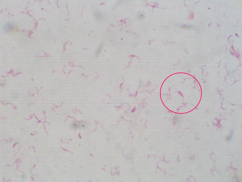
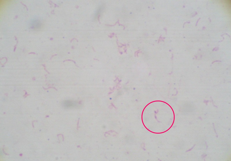
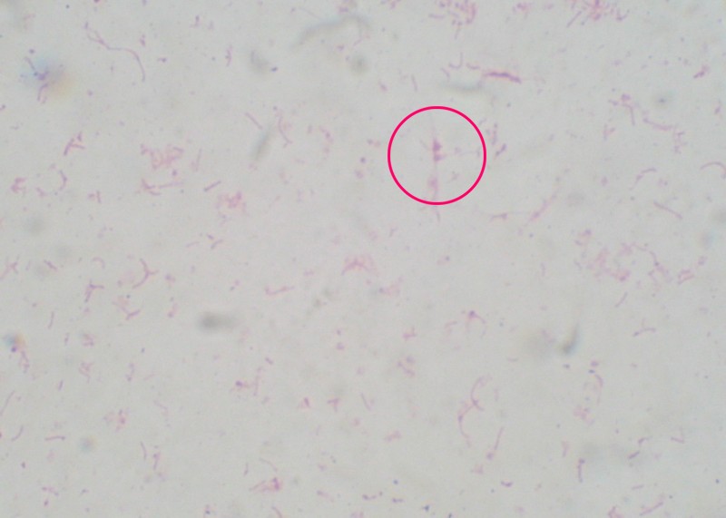
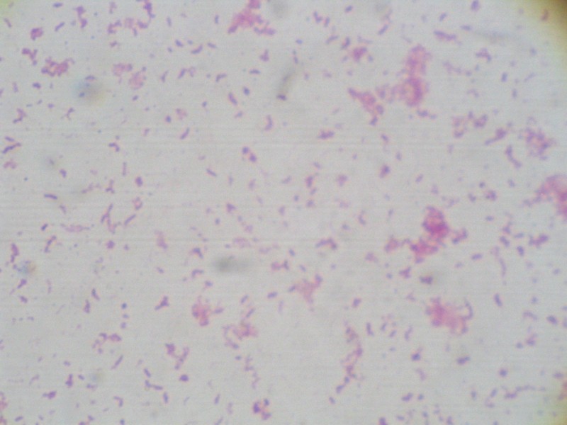
It's strange that the Helicobacter pylori cells have got different formations when grown on different media, and even on the same plate they may also look different:
I. Urea agar: Columbia blood agar base + 7% fetal bovin serum + HCl + urea + phenol red + antibiotics (vancomycin + amphotericin B + polymixin B)


II. CHBHP: Columbia blood agar base + 10% horse blood + antibiotics (vancomycin + amphotericin B + polymixin B)


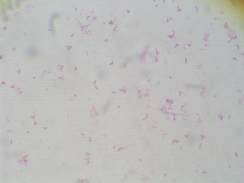
They're all gram negative so this time gram positive cocci have been removed (or suppressed), but as they've got several looks and some of them look different from what I know about helicobacters, now I can't really make sure if they are exactly Helicobacter... :(
 II. CHBHP: Columbia blood agar base + 10% horse blood + antibiotics (vancomycin + amphotericin B + polymixin B)
 They're all gram negative so this time gram positive cocci have been removed (or suppressed), but as they've got several looks and some of them look different from what I know about helicobacters, now I can't really make sure if they are exactly Helicobacter... :(
No comments:
Post a Comment